|
|
||
| HOME | ||
|
|
Training Program |
|
|
|
Contact Info |
|
|
|
Training Videos | |
|
|
Atlas of Ultrasound | |
|
|
Hands-on Classes | |
|
|
Bookings | |
|
|
||
|
|
||
|
|
||

|
FURTHER READING |
|||
|
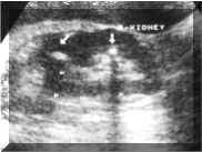
RENAL STONES Renal calculi are common and typically arise within the collecting system. Plain radiographs and intravenous urography are the traditional investigations for renal colic. Ultrasound has the advantage of demonstrating non-opaque calculi and hydronephrosis in comparison with the plain radiographs. Ultrasound has the potential of early diagnosis as compared with intravenous urography. Most renal calculi (about 80%) are calcified. Sonographically, stones usually appear as a hyperechoic foci with distal acoustic shadowing (Picture1). Gas may cause a similar appearance but may have a "dirty" shadow that is not as sharply defined as would occur with a calculus. There may be associated mucosal edema if the stone is impacted or if there is secondary inflammation or infection. Hydronephrosis and hydrourether may also be present. Tiny calculi may not cause distal shadowing if they are smaller than the focal zone of the transducer. When there is no shadowing, it may be difficult to distinguish small calculi from echogenic foci caused by fat or other echogenic reflectors within the renal sinus.
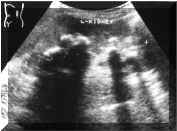
Ultrasound may have false-negative results with small calculi and suboptimal studies (e.g., obesity, uncooperative patient). Ultrasound may have false-positive results with renal parenchymal calcification, vascular calcification, and uretheric catheters. Staghorn calculi often appear as multiple, disconnected calculi within the collecting system (Picture2). Sonography generally underestimates the extent and size of stones in patients with staghorn and other large calculi. The presence of staghorn calculi may make it difficult to diagnose underlying hydronephrosis, because of strong acoustic shadowing from calculi.
Renal calcification also occur in patients with medullary sponge kidney and in patients with nephrocalcinosis. REFERENCES: |
|
||