|
|
||
| HOME | ||
|
|
Training Program |
|
|
|
Contact Info |
|
|
|
Training Videos | |
|
|
Atlas of Ultrasound | |
|
|
Hands-on Classes | |
|
|
Bookings | |
|
|
||
|
|
||
|
|
||

|
FURTHER READING |
|||
|
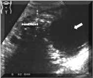
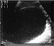
PANCREATIC PSEUDOCYSTS Pancreatic pseudocysts are differentiated from other other fluid collections largely on the basis of chronicity and the presence of mass effect. Acute pancreatic and peripancreatic fluid collections occur in more than half of patients with significant acute pancreatitis. They are nonencapsulated and usually lack significant mass effect, generally conforming to retroperitoneal and other fascial compartments. Sonographical Findings: Sonographically, pseudocysts are well defined and have variable internal echogenicity. Internal hyperechogenicity suggests hemorrhage. Pseudocysts may be septated or contain debris. A pseudocyst may be confidently diagnosed when a persistent fluid collection is detected in the clinical setting of pancreatitis. There is often mass effect on adjacent bowel loops and solid viscera, and occasionally a definable fibrous pseudocapsule (Pictures 1 and 2). Biliary obstruction may be caused by a pseudocyst. Percutaneous pseudocyst drainage may be successful.
Differential Diagnosis: Several pitfalls related to neoplasm should be kept in mind. Occasionally, a cystic pancreatic neoplasm may be mistaken for a pancreatic pseudocyst. The absence of clinical and laboratory findings of pancreatitis should prevent misdiagnosis. Conversely, a complex pseudocyst may stimulate a cystic neoplasm. REFERENCES: |
|
||