|
LIVER
ABSCESS
Ultrasound
is
the
most
commonly
used
imaging
technique
for
diagnosis
of
liver
abscess.
It
offers
a
high
sensitivity
and
specificity
in
diagnosis,
though
it
can
not
differentiate
pyogenic
from
amoebic
abscess.
Diagnosis
of
a
liver
abscess
is
easy
on
ultrasound
even
with
a
less
sophisticated
machine
and
a
less
experienced
person.
Careful
scanning
of
liver
is
essential
in
all
planes.
Patients
should
be
scanned
in
different
positions.
Decubitus
position
for
scanning
of
posterior
surface
of
liver
is
essential
to
pick
up
small
abscess
situated
posteriorly.
Presence
of
small
right
pleural
effusion
could
be
a
clue
to
underlying
abscess.
Most
often
sonography
is
diagnostic
and
no
other
imaging
technique
is
needed.
Ultrasound
is
useful
not
only
for
accurate
diagnosis
of
an
abscess
but
also
in
guiding
aspiration
if
required.
It
is
a
cheaper,
easier
and
reliable
technique
to
follow
up
patients.
Amoebic
Liver
Abscess
Amoebic
liver
abscesses
are
usually
single
but
can
be
multiple.
Typical
location
is
in
the
right
lobe
of
liver
subcapsular
close
to
the
diaphragm
and
posterolateral,
though
it
can
be
situated
in
any
location.
The
size
of
an
abscess
may
vary
from
few
centimeters
to
a
large
size
occupying
almost
entire
right
lobe
of
liver.
Very
early
stage
:
In
the
initial
stage,
cell
death
occurs
but
entire
dissolution
and
liquefaction
is
not
complete
as
the
contents
are
not
liquid.
This
may
be
termed
as
solid
abscess.
On
ultrasound
these
lesions
are
usually
small
and
probably
are
the
most
challenging
as
compared
to
the
other
stages
of
the
liver
abscesses.
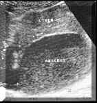
The
margins
of
the
abscess
may
be
ill
defined,
the
abscess
is
hypoechoic
as
compared
to
the
surrounding
liver.
However,
there
is
no
true
liquefaction
at
this
stage
and
therefore
there
is
poor
or
no
posterior
acoustic
enhancement.
The
demarcation
between
the
abscess
and
the
surrounding
liver
is
also
poor
(see
Picture1).

Picture1.
Early
abscess
[1].
At
this
stage,
the
differential
diagnosis
of
fat
spared
area
in
a
fatty
liver
or
an
early
neoplastic
lesion
have
to
be
considered.
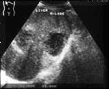
Recently
formed
amoebic
abscesses
:
An
abscess
of
recent
onset
has
a
distinct
central
liquified
area.
This
is
seen
on
ultrasound
as
a
sonolucent
or
an
hypoechoic
area
usually
with
fine
internal
echoes.
Because
of
the
liquefaction,
there
is
associated
posterior
acoustic
enhancement.
The
cavity
may
be
round,
oval
or
branching.
The
walls
of
the
abscess
at
this
stage
are
usually
not
very
thick
and
sometimes
the
demarcation
between
the
wall
and
the
surrounding
tissue
can
be
poor.
Sometimes
the
walls
may
be
thicker
and
these
may
be
seen
as
shaggy,
ill-defined
echogenic
areas
along
the
walls
(see
Picture2).
It
is
at
this
stage
of
the
abscess
that
aspiration
may
be
required.
Small
amount
of
air
in
the
abscess
because
of
secondary
infection
or
following
an
aspiration
is
seen
as
highly
reflective
dots.
Picture2.
It
is
at
this
stage
of
the
abscess
that
differential
diagnosis
of
a
cyst
in
the
liver,
a
cyst
with
haemorrhage,
cystic
metastatic
deposit
or
sometimes
a
hydatid
cyst
and
haematoma
are
to
be
considered.
Abscesses
of
some
duration
:
The
basic
difference
between
an
acute
abscess
and
an
abscess
of
some
duration
is
that,
in
the
latter
the
body
has
had
time
to
wall
up
the
lesion
by
producing
a
layer
of
fibrous
tissue
around
it.
On
sonography
an
abscess
shows
thick
walls
which
may
vary
from
a
few
mm
to
1.5
cm
in
thickness.
The
echogenicity
of
the
abscess
also
varies,
abscesses
generally
become
more
sonolucent
at
this
stage,
some
abscesses
become
more
echogenic
because
of
organisation
of
fluid
(see
Picture3).
Picture3.
Healing
Stage
:
The
abscess
heals,
the
liquid
contents
dry
up,
which
has
been
described
as
putty
appearance.
On
ultrasound
it
is
seen
again
as
a
lesion
with
thick
walls
fairly
echogenic
as
compared
to
surrounding
organs.
This
shadow
can
be
seen
on
ultrasound
for
a
long
time,
even
years.
It
is
usually
at
this
stage
that
the
differential
diagnosis
of
a
neoplasm,
haemangioma
or
granuloma
in
liver
come
into
picture.
Summary:
The
diagnosis
of
liver
abscess
is
easy
on
ultrasound
and
besides
pointing
out
the
diagnosis,
the
number
of
abscesses
and
helping
in
aspiration,
if
the
sonologist
can
predict
the
stage
of
evolution
then
it
could
help
a
clinician
in
deciding
the
management
of
a
patient.
REFERENCE:
[1]
N.G.
CHAUBAL,
"Follow
up
of
Amoebic
Liver
Abscess
with
Ultrasound
and
the
Role
of
New
Techniques
in
Ultrasound
Including
Colour
Doppler.",
www.bhj.org/journal/oct97. |
