|
|
||
| HOME | ||
|
|
Training Program |
|
|
|
Contact Info |
|
|
|
Training Videos | |
|
|
Atlas of Ultrasound | |
|
|
Hands-on Classes | |
|
|
Bookings | |
|
|
||
|
|
||
|
|
||

|
FURTHER READING |
|||
|
GESTATIONAL
TROPHOBLASTIC
NEOPLASIA Benign GTN Complete
Hydatidiform
Mole: Diagnosis
in
First
Trimester: Diagnosis
in
Second
Trimester:
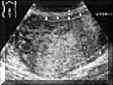
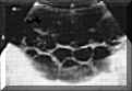
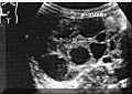
Theca-lutein cysts are the ovarian response to excess human chorionic gonadotropin (hCG) secretion by GTNs. These cysts are formed by large follicles that are full of liquid and have smooth walls, and that partially or totally occupy the ovaries (Picture2). Although they may be unilateral, it is more common to find them bilaterally. Theca-lutein cysts appear only with total hydatidiform moles, and they may persist in postmolar disease.
Approximately 2 percent of all molar pregnancies have a coexisting fetus although such a fetus is seldom alive. Partial
Hydatidiform
Mole: When a pregnancy is more than 12 weeks and the fetus is intact, it is not difficult to diagnose a partial mole because the typical "snowflake" picture is seen in the placental area accompanying the fetus. Nevertheless, this case is not typical, as the embryo usually degenerates early and is resorbed. As the vesicular degeneration is not complete, or does not occur as early as for the total hydatidiform mole, the picture is not as clear as it is for the total mole. Distinction between the partial hydatidiform mole and the molar pregnancy with coexistent fetus is important as they differ in malignant potential and karyotype. A partial mole is considered to have less malignant potential. Differential
Ultrasound
Diagnosis: 1- Missed abortion: The picture most easily confused with a postmolar GTN is a spontaneous abortion because what is seen are echo-refringent and non-homogeneous chorionic remains either located inside the cavity or attached to the uterine wall. Low or negative hCG levels are helpful for differentiating these entities. 2- Blighted ovum (degenerated ova): It can be confused with partial mole. The main difference between a partial mole and blighted ovum lies in the perfect interior delimitation of the embryonic sac in the latter. The partial mole usually has a poorly defined sac surrounded by a strongly sonolucent, trophoblastic ring; it shows remains of an embryo, which never appears in a blighted ovum. 3- Ectopic pregnancy: In ectopic pregnancy a decidual picture may be seen similar to that of a mole because it shows pseudovesicles and a pseudosac. The combined use of quantitative determinations of hCG and vaginal ultrasound may resolve this uncertainty. 4- Hydropic placental degeneration: It can be confused with a mole accompanying a live fetus. Vesicles, cysts, fetal remains, and an abnormal placenta can be seen. The clinical history of the patient - including the possibilities of diabetes, isoimmunization, and intragestational infection - should be considered carefully. 5- Leiomyoma: It may be confused with a total hydatidiform mole. Leiomyomas have a characteristic whorling and lack the cystic appearance of a mole. Shadowing from echodense areas is found in degenerated leiomyomas 6- Retained products of conception: Low levels of hCG are generally found. 7- Ovarian tumors: They may be diagnosed when a normal uterus is demonstrated. Malignant GTN The malignant variants of trophoblastic disease are invasive mole and choriocarcinoma. Sonographic detection of invasion of myometrium is an indication of malignancy. Sonography is part of the routine recommended investigation for the staging of gestational trophoblastic neoplasia. The pelvis and liver are scanned to establish the extent of the pelvic disease and to search for metastases. REFERENCE: |
|
||