|
CHRONIC
PANCREATITIS
Chronic
pancreatitis
is
defined
as
a
prolonged
inflammatory
disease
of
the
gland
associated
with
progressive
parenchymal
destruction
and
loss
of
endocrine
and
exocrine
functions.
Ultrasound
Findings
Image
findings
are
generally
poor
predictors
of
the
clinical
severity
of
chronic
pancreatitis.
Multiple
Calcifications:
The
calcifications
are
caused
by
stones
within
the
main
or
branch
pancreatic
ducts.
Detecting
multiple
pancreatic
calcifications
allows
a
confident
diagnosis
of
chronic
pancreatitis.
Sonography
is
less
sensitive
than
CT
in
detecting
pancreatic
calcifications.
On
occasion,
a
diffusely
hyperechoic
pancreas
may
be
an
indicator
of
chronic
pancreatitis.
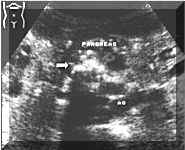
Picture1.
Multiple
pancreatic
calcifications
(see
arrow).
Ductal
Dilatation:
Chronic
pancreatitis
may
cause
dilatation
of
the
pancreatic
or
common
bile
duct.
Some
researchers
feel
that
dilatation
of
the
main
pancreatic
duct
is
the
most
reliable
sign
of
chronic
pancreatitis.
Focal
Mass:
A
focal
mass
is
seen
in
about
one
third
of
patients
with
chronic
pancreatitis.
This
results
from
proliferation
of
fibrous
tissue
and
infiltration
by
inflammatory
cells.
Sonographically,
the
mass
is
usually
small,
measuring
about
2
to
3
cm
in
diameter.
It
is
usually
hypoechoic,
with
a
disorganized
echo
pattern.
However,
there
is
a
wide
range
in
size
and
texture.
Some
masses
may
be
quite
large
and
strongly
echogenic.
Focal
masses
with
chronic
pancreatitis
must
be
differentiated
from
neoplasm.
A
few
features
favor
the
diagnosis
of
a
benign
inflammatory
mass
over
a
tumor:
1-Calcification
within
the
mass
makes
the
diagnosis
of
chronic
pancreatitis
almost
certain.
Hyperechoic
masses,
even
those
without
discrete
calcifications,
are
usually
caused
by
chronic
pancreatitis,
even
though
hyperechoic
carcinoma
is
not
rare.
2- The
presence
of
duct
dilatation
within
the
mass
generally
means
that
the
mass
is
caused
by
chronic
pancreatitis,
although
carcinoma
may
occasionally
have
internal
hypoechoic
regions
as
well.
Pseudocysts:
Chronic
asymptomatic
pseudocysts
may
be
found
in
patients
with
chronic
pancreatitis.
Pseudocysts
also
occur
in
association
with
pancreatic
carcinoma.
Summary
The
role
of
ultrasound:
1-
to
document
the
morphological
abnormalities
of
the
gland.
These
include
focal
mass,
ductal
dilatation,
and
stones
2-
to
monitor
the
development
and
evolution
of
pseudocysts
3-
to
look
for
associated
abnormalities
that
may
occur
elsewhere
in
the
abdomen.
REFERENCES:
[1]Abdominal
Ultrasound.
E.E.Sauerbrei,
K.T.Nguyen,
R.L.Nolan.
1992.
2]Sonography
of
the
Abdomen.
R.B.Jeffrey,
P.W.Rolls.
1995
|
